
Figure 4. Quantitative investigation of the influence of perivascular space (PVS) on DTI, across 861 subjects of the human connectome project data.
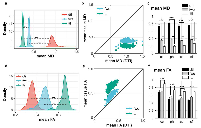
Values from DTI were compared with those from free water elimination (FWE) and tissue tensor imaging (TTI) techniques. Mean MD of the PVS voxels are compared in (a) and (b). Mean MD values were compared across four white matter regions from Desikan-Killiany atlas (c). Regions are: corpus callosum (cc), para-hippocampus (ph), centrum semiovale (cs) and superior-frontal part of the white matter (sf). All differences are corroborating simulation and subject-level results and are significant at p<0.001 (using paired t-test). Similar investigation on the fractional anisotropy (FA) values are shown in (d-f). Diffusivity values are in μm2/ms.