
Figure 1. Exposure of cardiac allografts to circulating mitochondria increases costimulation blockade resistant rejection.
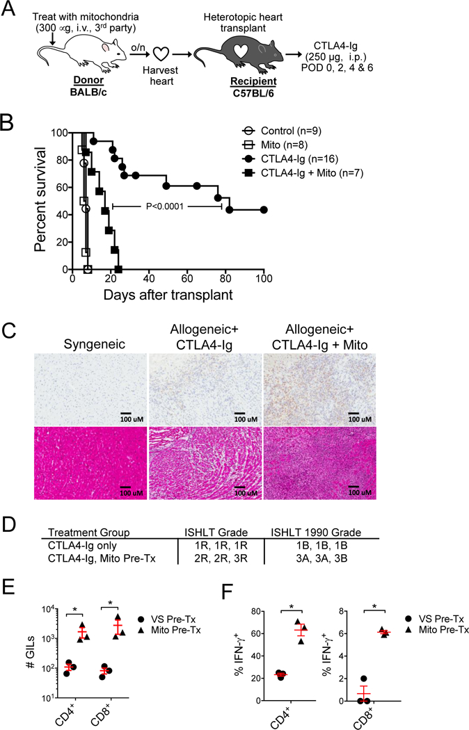
A) Donor hearts were harvested from BALB/c mice (H-2d) following intravenous injection of mitochondria (300 mg) or vehicle solution (control) one day prior. Hearts where then heterotopically transplanted into C57BL/6 mice (H-2b) treated with or without costimulation blockade with murine CTLA4-Ig (250 mg, i.p., POD 0, 2, 4, 6). B) Kaplan-Meier analysis of cardiac allograft survival. Curves were compared by log-rank test. C) CD3 immunohistology (top panels) and H&E staining (lower panels) of 5 mm cryosections from syngeneic or allogeneic cardiac allografts at 2-weeks post-transplantation. Grafts were from donors exposed to mitochondria (Mito) or vehicle (control) one day prior to organ harvest. Allogeneic recipients were treated with CTLA4-Ig. D) Current and 1990 ISHLT histologic grading of cardiac allograft rejection in the allogeneic transplant groups (n=3 per group). E) Quantification of graft-infiltrating CD4+ and CD8+ T cells. Graft infiltrating leukocytes (GILs) were purified from cardiac allografts harvested 2-weeks post-transplantation and assayed by flow cytometry for CD4+ and CD8+ T cells. Allogeneic cardiac allografts were harvested from donor mice exposed to circulating mitochondria (Mito Pre-Tx, n=3) or treated with vehicle solution (VS, n=3) one day prior. F) Percentage of CD4+ and CD8+ GILs producing IFN-g in cardiac allografts in from donors pre-treated with intravenous mitochondria (Mito Pre-Tx, n=3) or VS (n=3). Comparisons were performed using a two-tailed Student’s t-test. *A p-value <0.05 was considered significant.