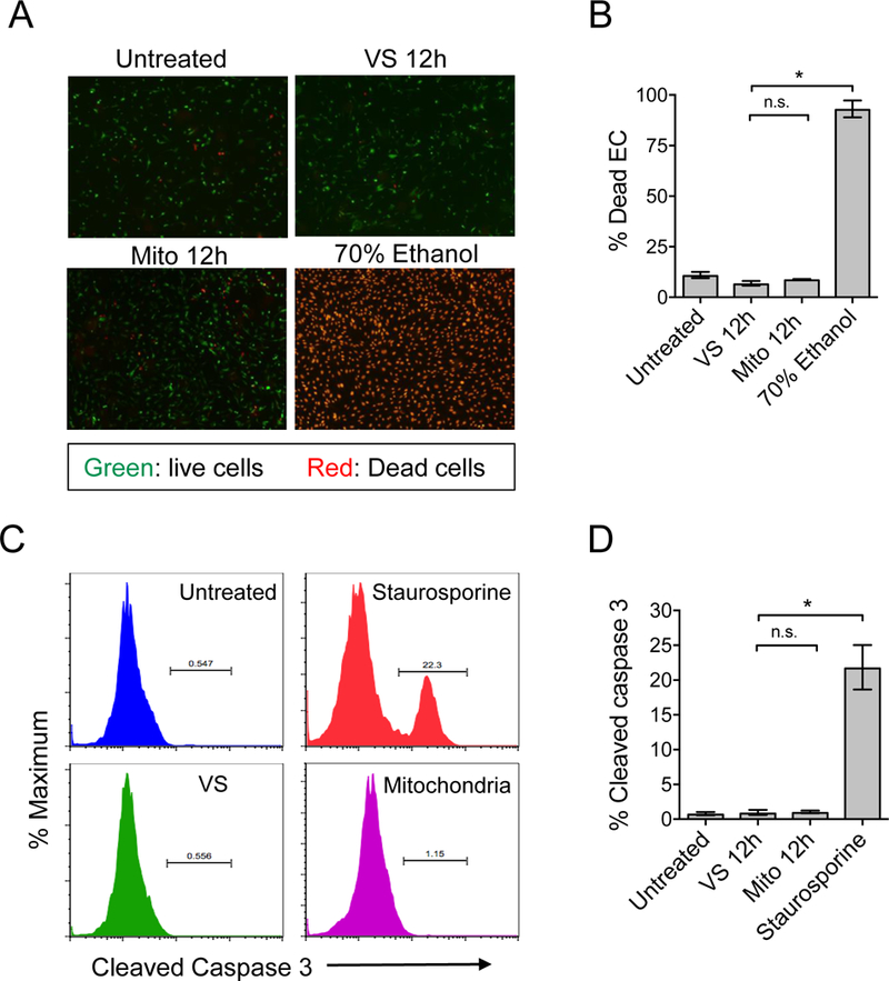
Figure 7. Determination of EC viability and apoptosis following mitochondrion–EC interaction.

A) The viability of mitochondria-stimulated ECs was evaluated by staining with live/dead cell viability dye. Live cells stain green and dead cells stain red in this assay. ECs treated with staurosporine were used as positive controls. B) No significant increase of cell death was revealed at 12 hours post mitochondrion–EC interaction. C) Detection of EC apoptosis during mitochondrion–EC interaction was determined by detection of cleaved caspase 3. A representative experiment of ECs treated with mitochondria, vehicle solution (VS), or staurosporine (positive control). Following their interaction with exogenous mitochondria, ECs did not undergo significant apoptosis.
