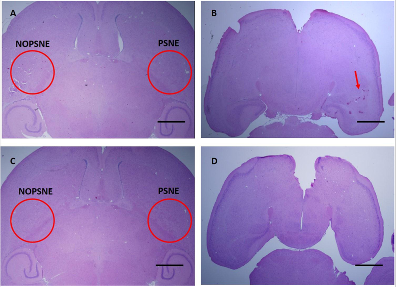
Figure 9:
Examples of H&E staining from two subjects that were injected with non-diluted PSNE and sonicated at 1.5 MPa. (A) The slice that was located 3 mm from the bottom of the brain from subject 1. No apparent damage was created by sonication within the targeted area with or without PSNE. (B) H&E slice from the bottom of the brain from subject 1. Red arrow is pointing at the side that was sonicated with PSNE. Some micro-hemorrhage was observed, but it was not as extensive as subjects sonicated at a higher pressure. This was observed only in 1 of the 4 mice. (C) H&E slice that was located 3 mm from the bottom of the brain from subject 2. No apparent damage was found in both side. (D) H&E slice from the bottom of the brain from subject 2. There was no apparent damage at the bottom in both side. Scale bars are 1mm.

