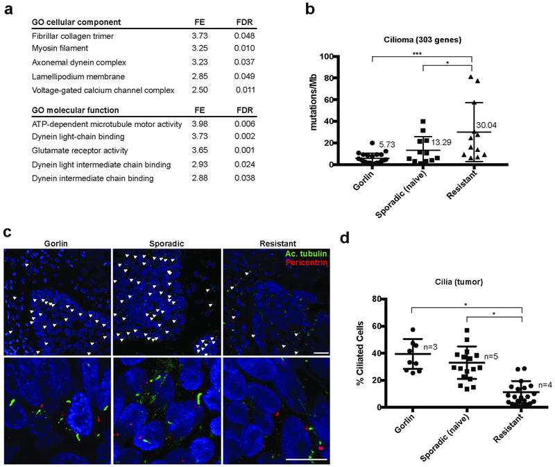
Figure 1. Human resistant BCC harbor reduced primary cilia.
(a) Top-lists of GO cellular components and molecular functions associated with genes commonly mutated in resistant BCCs.
(b) Quantification of the mutations found in the ciliome of Gorlin patients, sporadic naïve and resistant human BCCs using whole exome sequencing. Numbers indicate the mean values for each category.
(c) Acetylated tubulin (cilia shaft), pericentrin (cilia body) and DAPI immunostainings of Gorlin patient, sporadic naïve and resistant human BCCs. White arrowheads indicate cilia.
(d) Quantification of cilia in the tumor compartment shown in (c). Dots represent microscope fields, distributed across n>3 tumors.
FE stands for fold enrichment. FDR stands for false-discovery rate. Scale bars indicate 25μm. Horizontal bars and error bars in (b) and (d) represent the mean ± SD. *p < 0.05, ***p < 0.001.

