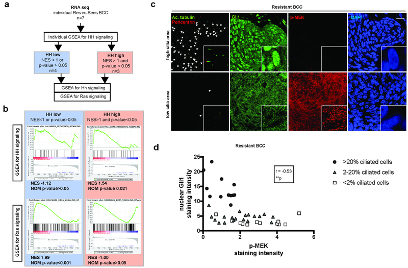
Figure 2. Loss of primary cilia goes with HH pathway inactivation and Ras/MAPK pathway activation in a subset of resistant BCCs.
(a) Algorithm for the analysis of RNA-sequencing obtained from vismodegib-sensitive and - resistant human BCCs.
(b) GSEA for HH and Ras pathways activation in “HH-high” versus “HH-low” human resistant BCCs. NES stands for normalized enrichment score.
(c) Adjacent immunostainings for cilia (acetylated tubulin and pericentrin), Gli1 (as a readout for HH pathway activation) and p-MEK (as readout for Ras/MAPK pathway activation) in resistant human BCCs. White arrowheads indicate cilia. Higher magnifications are shown in framed pictures.
(d) Correlation between nuclear Gli1 and p-MEK relative intensity in resistant human BCCs, annotated for cilia density. Dots represent microscope fields, distributed across 4 different resistant BCC tumors.
Scale bars indicate 25μm. **p < 0.01. r stands for Spearman correlation factor.

