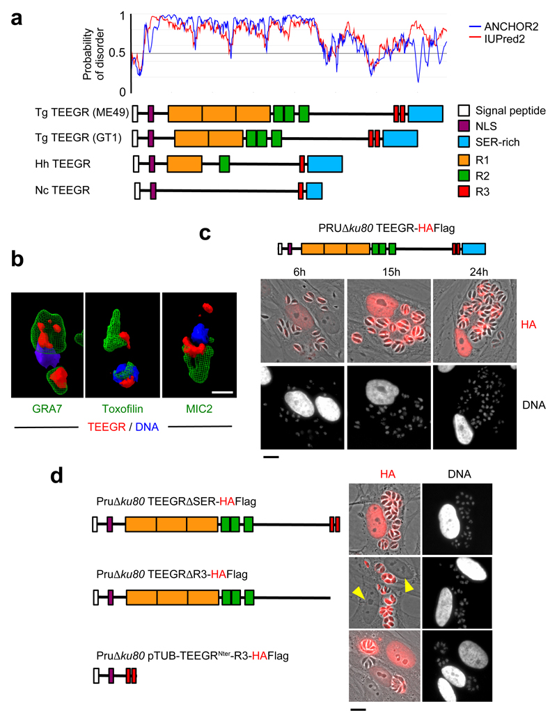
Fig. 1. The export of TEEGR in the host cell nucleus.
The gene TGME49_239010 encoding TEEGR was originally identified in silico together with GRA164, GRA245 and TgIST6,7 as strong parasite candidate genes with attributes to both target the parasite secretory pathway and to reach the host cell nuclei. a, TEEGR is a highly disordered 80 kDa protein (predicted disorder score > 0.5) that carries a signal peptide, a putative nuclear localization sequence (NLS) and multiple repeated domains (R1, R2 and R3) whose repeat number and distribution have evolved through coccidian subclasses and among T. gondii lineages. Tg: Toxoplasma gondii, Nc: Neospora caninum, Hh: Hammondia hammondia. b, HAFlag (HF)-tagged TEEGR (in red) in Pru∆ku80 extracellular parasites is contained in cytoplasmic organelles distinct from the apical micronemes (MIC2, in green) and rhoptries (Toxofilin, in green), and partially co-localizing with the dense granule protein GRA7 (in green). Cells were co-stained with Hoechst DNA-specific dye (in blue). Scale bars, 1 μm. c, Time course of TEEGR secretion and export to the host cell nucleus. Human Foreskin Fibroblasts (HFF) were infected with a Pru∆ku80 TEEGR-HF and stained with anti-HA antibodies (red). Scale bars, 10 μm. d, in situ subcellular localization of TEEGR chimeric proteins (in red) in HFF infected with parasites expressing endogenously HF-tagged TEEGR truncated proteins (TEEGR∆SER and TEEGR∆R3) or TEEGRNter-R3 expressed under the control of a TUB8 promoter. Scale bars, 10 μm. All data are representative of 3 independent biological experiments.

