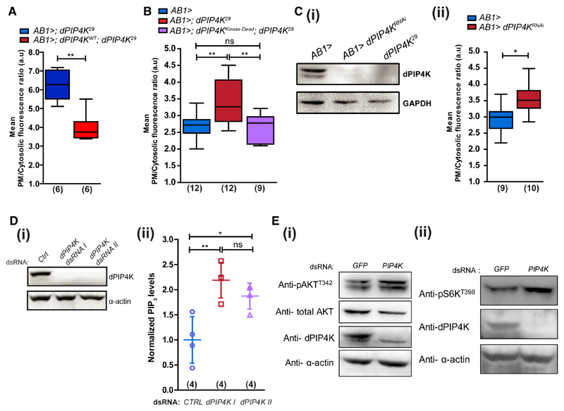
Figure 2. dPIP4K Cell-Autonomously Controls PIP3 Levels.
PIP3 levels in salivary glands.
(A) Mutant and wild-type dPIP4K rescue.
(B) Control, mutant, and kinase-dead dPIP4K rescue.
(C) (i) Immunoblot (from wandering third instar larvae) showing salivary gland-specific depletion of dPIP4K (dPIP4KRNAi). (ii) PIP3 levels in control and dPIP4KRNAi salivary glands.
(D) (i) Immunoblot for dPIP4K knockdown in S2R+ cells with two different dsRNAs. (ii) Total PIP3 using LCMS in whole cell lipid extracts of S2R+ cells treated with indicated dsRNAs (data pooled from two experiments, a total of four biological replicates). Scatter plots with mean ± SD. Statistical test: one-way ANOVA with post hoc Tukey’s multiple pairwise comparison. *p value < 0.05; **p value <0.01.
(E) Western blots for (i) pAKTT342 and (ii) pS6KT398 levels in control and dPIP4K knockdown cells.
See also Figure S2.

