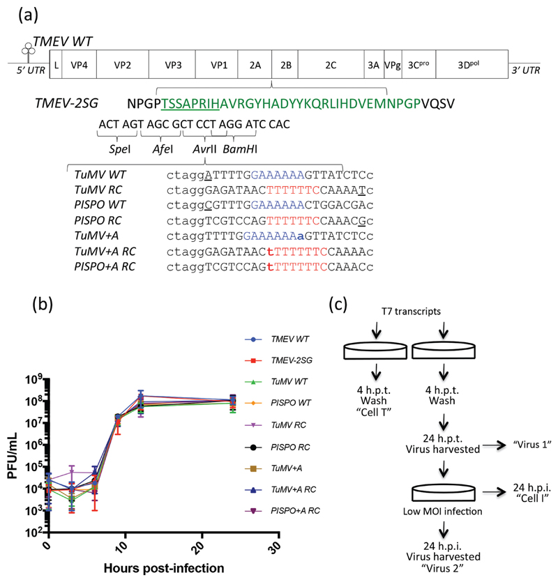
Fig. 2.
Introduction of duplicate StopGo and potyvirus-derived slip-site sequences do not affect TMEV growth kinetics. (a) Schematic of the TMEV WT genome, indicating the duplicate 2A StopGo peptide (green) inserted to create TMEV-2SG. Underlined residues indicate those encoding cloning sites (detailed underneath) that do not contribute to StopGo function. Wild-type flanking residues are shown in black. Seven sequences based on potyvirus polymerase slippage sites were inserted into TMEV-2SG. Slip sites are shown in blue (native potyviral slip sites and ‘+A’ variants) or red (reverse complements); additional nucleotides extending the slip site are indicated in bold; other sequences have an additional nucleotide (underlined) added at the end of the insert to maintain the same reading frame (this is checked in each read to guard against inter-sample contamination from the +A mutants as a result of multiplex tag misassignments). Upper case indicates nucleotides derived from potyvirus genomic sequences; lower case indicates flanking nucleotides used for cloning. (b) One-step growth curves (n=2) of mutant viruses indicate they do not exhibit significantly altered growth kinetics compared to either TMEV WT or TMEV-2SG. (c) Schematic of the experimental protocol followed to obtain the initially transfected cells (‘Cell T’), first-round virus (‘Virus 1’), infected cells (‘Cell I’) and second-round virus (‘Virus 2’) samples.

