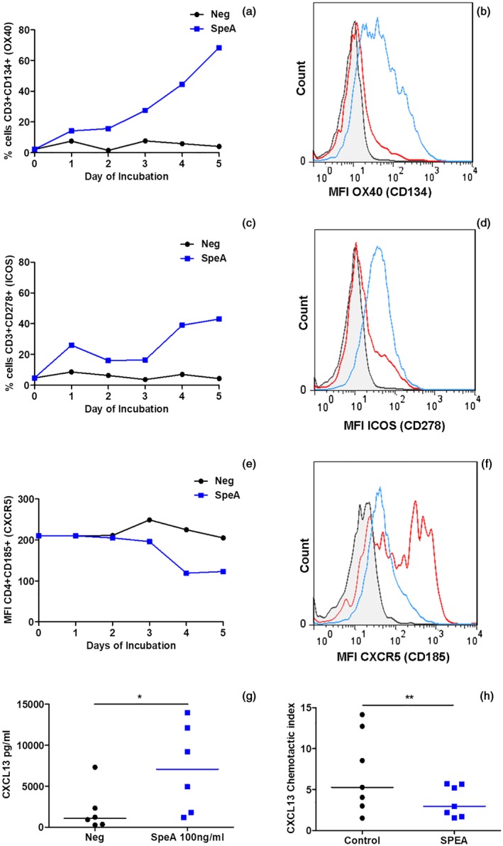
Figure 6.

Altered T follicular helper (Tfh) phenotype in streptococcal pyrogenic exotoxin (SpeA)‐stimulated tonsil cells. Tonsil cells stimulated with SpeA (blue squares) showed a marked early increase in the percentage and mean fluorescence intensity (MFI) of CD3+ T cells expressing OX40 (CD134, a,b, P = 0·029) and inducible T cell co‐stimulator (ICOS) (CD278, c,d (P = 0·029) compared with unstimulated controls (black circles) (Wilcoxon’s paired signed‐rank t‐test). Four tonsil donors for OX40 and ICOS, representative time–course (days 1–5) and fluorescence activated cell sorter (FACS) plot of gated T lymphocytes are shown from one donor each (day 5); grey‐shaded, isotype control; red line, unstimulated cells; blue line, SpeA 100 ng/ml. In contrast, expression of C‐X‐C motif chemokine receptor (CXCR5) in CD4+ cells reduced in response to SpeA compared with unstimulated controls (e,f, P = 0·015*, Wilcoxon’s paired signed‐rank t‐test). Seven donors for CXCR5, time–course and FACS plot from one representative donor (day 5) as described above. SpeA stimulated tonsil cells showed an increase in chemokine (C‐X‐C motif) ligand (CXCL)13 production (g, P = 0·0313 Wilcoxon’s paired signed‐rank t‐test) but chemotaxis of tonsil CD4+CXCR5+ cells toward CXCL13 gradient was reduced by prior co‐incubation with SpeA [h, P = 0·005**, two way analysis of variance (anova)].
