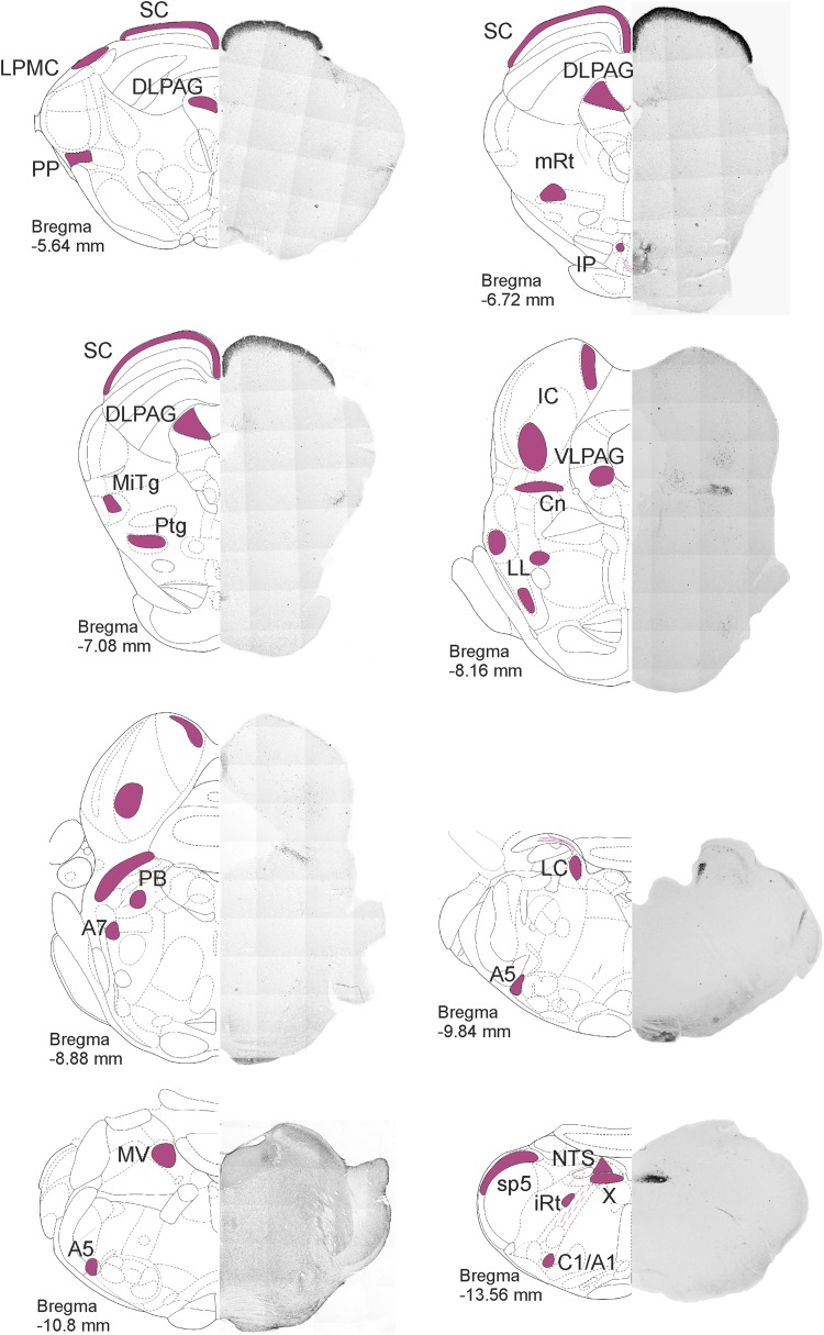
Fig. 1.
Distribution of secretagogin in the rat brainstem I. Secretagogin+ somata were identified in coronal sections throughout the rat brainstem. Low-power micrographs are paired with schemata of coronal brain sections to indicate craniocaudal levels. Secretagogin+ neurons-containing regions are indicated in purple. Superior colliculus, the microcellular tegmental nucleus, the dorsal nucleus of vagus and the noradrenergic cell groups including A1, A5, A6 and A7 typically expressed secretagogin. For high-power images, please see Fig. 4. A1, A5, A7 noradrenergic cell groups, C1 adrenergic cell group, Cn cuneiform nucleus, DLPAG dorsolateral periaqueductal grey, IC inferior colliculus, IP interpeduncular nucleus, iRT intermediate reticular nucleus, LC locus coeruleus, LL lateral lemniscus, LPMC lateral posterior thalamic nucleus mediocaudal part, MiTg microcellular tegmental nucleus, mRT mesencephalic reticular formation, MV medial vestibular nucleus, NTS solitary tract nucleus, PB parabrachial nucleus, PP peripeduncular nucleus, Ptg pedunculopontine tegmental nucleus, sB subbrachial nucleus, SC superior colliculus, sp5 spinal tract nucleus of V, VLPAG ventrolateral periaqueductal grey, X dorsal nucleus of X

