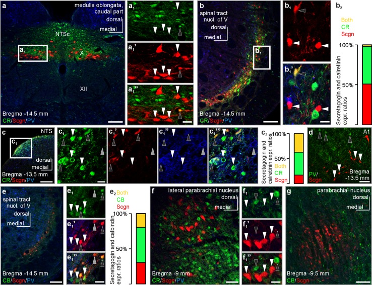
Fig. 5.
Co-expression of secretagogin with classical Ca2+-binding proteins in select brainstem nuclei of the rat. a–a1’’ Complementary distribution of calretinin and secretagogin in the commissural part of the solitary nucleus and the dorsal nucleus of vagus (white arrowheads indicate secretagogin+/calretinin− neurons, black arrowhead points to a secretagogin+/calretinin+ neuron). b–b1’ Secretagogin+ neurons typically remained calretinin immunonegative (white arrowheads) in the spinal tract nucleus of the trigeminus, with exceptional co-expression only (black arrowhead). b2 Single- and co-expression ratios of secretagogin and calretinin in the spinal tract nucleus of the trigeminus. 100% percentage refers to all labelled cells detected for secretagogin and/or calretinin immunoreactivity. Secretagogin+/calretinin+ neurons: 2.8 ± 3.2%, secretagogin+/calretinin− neurons 51.2 ± 6.1%, secretagogin−/calretinin+ neurons 47.5 ± 5.4% (as of average ± s.e.m.). c–c1’’’ Calretinin+ neurons in the dorsolateral part of the solitary tract nucleus can either remain immunonegative for secretagogin (white arrowheads) or co-express it (black arrowheads) Grey arrowhead points to a CR−/Scgn−/PV+ neuron. c2 Single- and co-expression ratios of secretagogin and calretinin in the dorsolateral part of the solitary tract nucleus. 100% percentage refers to all labelled cells detected for secretagogin and/or calretinin immunoreactivity. Secretagogin+/calretinin+ neurons: 36.1 ± 2.3%, secretagogin+/calretinin− neurons 16.9 ± 3.5%, secretagogin−/calretinin+ neurons 43.7 ± 2.1% (as of average ± s.e.m.). d Secretagogin+ neurons (white arrowheads) in the A1 field did not co-express parvalbumin (black arrowheads point to secretagogin−/parvalbumin+ somata). e–e1’’ Secretagogin+ neurons typically co-expressed calbindin in the spinal tract nucleus of the trigeminus (black arrowheads). White arrowheads point to calbindin+/secretagogin− somata, grey arrowhead to a secretagogin+/calbindin− soma. e2 Single- and co-expression ratios of secretagogin and calbindin in the spinal tract nucleus of the trigeminus. 100% percentage refers to all labelled cells detected for secretagogin and/or calbindin immunoreactivity. Secretagogin+/calbindin+ neurons: 19.1 ± 8.0%, secretagogin+/calbindin− neurons 30.7 ± 11.3%, secretagogin−/calbindin+ neurons 50.2 ± 7.2% (as of average ± s.e.m.). f–f1’’ Secretagogin+ neurons showed a complementary distribution to calretinin+ neurons in the lateral parabrachial nucleus (white arrowheads point to secretagogin+/calretinin− somata, black arrowheads point to secretagogin−/calretinin+ somata). g Secretagogin+ neurons remain immunonegative for calbindin in the parabrachial nucleus. CB calbindin, CR calretinin, NTS solitary tract nucleus, NTSc commissural part of the solitary tract nucleus, Scgn secretagogin, PV parvalbumin, X dorsal nucleus of vagus, XII hypoglossal nucleus. Scale bars 150 µm (a, b, c, e), 70 µm (g), 40 µm (d, f), 10 µm (a1’’, b1’, c1, e1’’, f1’’)

