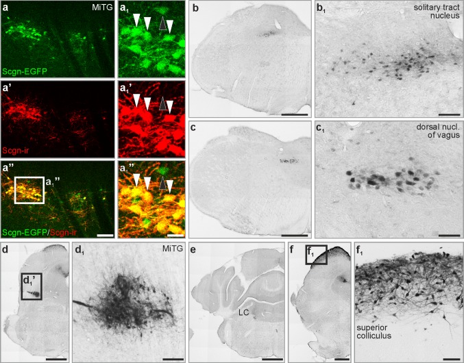
Fig. 6.
Secretagogin expression in the mouse brain stem. a–a1’’ EGFP-expressing neurons in secretagogin–EGFP mice showed secretagogin immunoreactivity as exemplified in the microcellular tegmental nucleus (white arrowheads in a–a1’’). Occasionally, EGFP-expressing neurons remained secretagogin immunonegative (black arrowhead in a–a1’’). b–f1 Secretagogin+ neurons in the solitary tract nucleus (b, b1), dorsal nucleus of vagus (c, c1), microcellular tegmental nucleus (d, d1), locus coeruleus (e) and the superior colliculus (f, f1). LC locus coeruleus, MiTg microcellular tegmental nucleus. Scale bars 1 mm (b–f), 100 µm (a’’, d1, f1), 70 µm (b1), 40 µm (c1)

