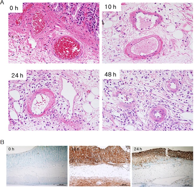
Figure 4.
Histological change over time in the stomach after IRE (haematoxylin & eosin staining and TUNEL assay). (A) Immediately after IRE, there were no remarkable changes except for subtle mucosal depression and haemorrhage. At 10 h after IRE application, tissue damage was noted in the electroporated area. At 24 and 48 h after IRE application, tissue damage in the electroporated area and neutrophil infiltration throughout the treated area were observed. (B) Vigorous apoptosis was observed at 10 h after IRE which correlated with H&E staining results.

