Figure 3.
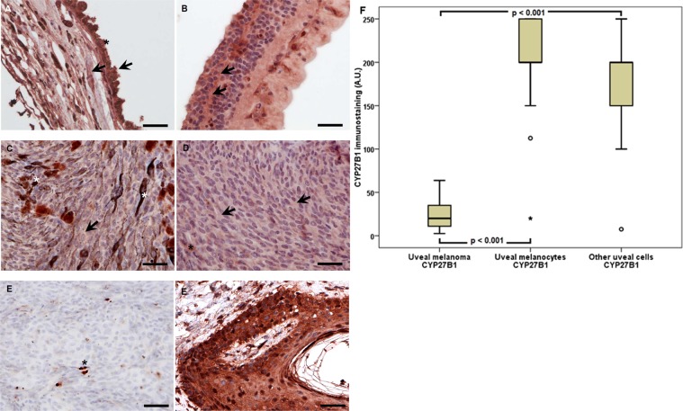
Representative CYP27B1 immunostaining in uveal pigmented cells (A), other cells (B), strongly pigmented (C) and slightly pigmented (D) melanomas and negative (E) and positive (F) controls. Arrows indicate CYP27B1 immunostaining, asterisks indicate melanin, scale bar: 50 μm. Mean CYP27B1 immunolabelling in melanoma, normal melanocytes and other normal uveal cells (F) (statistically significant differences are denoted with the Wilcoxon signed-rank test).

