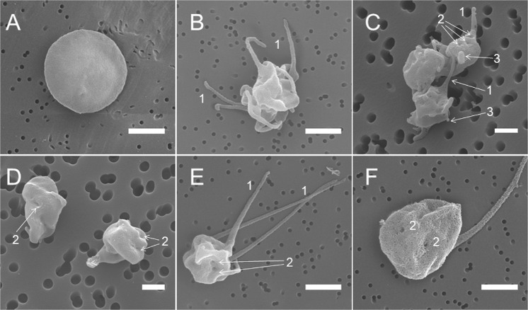
Fig. 2. Representative scanning electron micrographs of platelets under various experimental conditions.
a A control untreated (resting) platelet; b a platelet treated with KKO/PF4; c an aggregate of platelets treated with KKO alone; d platelets treated with PF4 alone; and e, f a platelet treated with A23187. All the platelet samples were incubated for 15 min at 37 °C. Final concentrations: 10 µg/ml PF4, 50 µg/ml KKO, and 12 µM Ca2+-ionophore. Arrows and numbers indicate 1—filopodia/pseudopodia, 2—pores of the open canalicular system, and 3—blebs and knobs. Magnification bars: 1 µm

