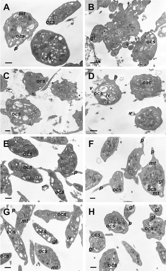
Fig. 4. Representative transmission electron micrographs of platelets under various experimental conditions.
a Control untreated resting platelets, b platelets treated with Ca2+-ionophore A23187, c, d platelets treated with KKO/PF4 for 15 min (c) and 60 min (d), e, f platelets treated with KKO alone for 15 min (e) and 60 min (f), g, h platelets treated with PF4 alone for 15 min (g) and 60 min (h). Final concentrations: 10 µg/ml PF4, 50 µg/ml KKO, and 12 µM Ca2+-ionophore. Designations: α, α-granules; gl, glycogen granules; lv, lytic vacuole; m, mitochondria; mt, microtubules; ocs, open canalicular system; p, pseudopodium; v, microvesicles. Magnification bars: 0.5 μm. KKO/PF4 complexes (c, d) induce profound ultrastructural changes in platelets similar to those observed in the positive control (b), while platelets treated with KKO and PF4 alone (e–h) remain largely unperturbed, as in the negative control (a)

