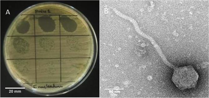
Figure 1.
(A) Bacteriophage FNU1 spotted onto F. nucleatum culture on BHI with 1% agar. From the top left to the bottom right, each square represents a 10-fold serial dilution of FNU1. Clearing is seen at the highest concentrations with individual plaques discernible at subsequent dilutions. Each single plaque is ≈1 mm diameter. (B) TEM image of FNU1 revealing Siphoviridae bacteriophage with long ≈310 nm tail and icosahedral head diameter of ≈88 nm.

