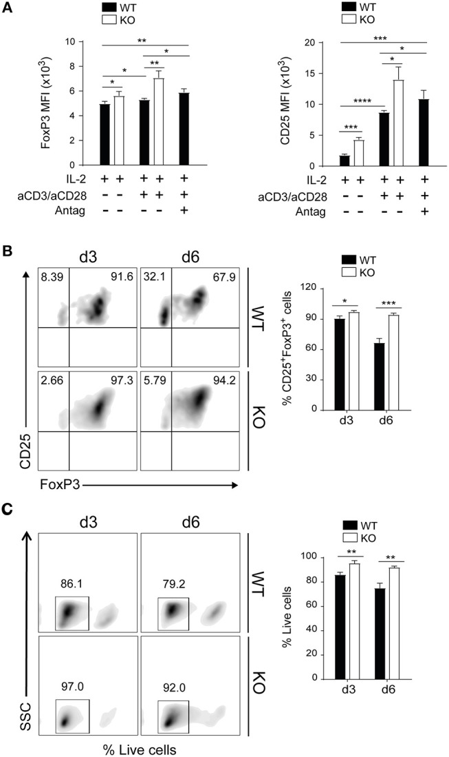
Figure 2.

PAR4 signaling regulates stability and survival of Foxp3+ Tregs in vitro. CD4+CD25+ Tregs were freshly isolated from spleen and pLNs of WT (filled bars) and PAR4ko (open bars) mice. The Tregs were activated with anti-CD3/anti-CD28 in the presence of IL-2 for 3 days and in some cases PAR4 antagonist (Antag) was added to WT Tregs. The MFI levels of FoxP3 and CD25 expression were analyzed by flow cytometry (A). Flow cytometry assessment of FoxP3 and CD25 expression on Tregs activated by anti-CD3/anti-CD28 in presence of IL-2 for 3 and 6 days (B). Flow cytometry assessment of live Tregs activated by anti-CD3/anti-CD28 in presence of IL-2 for 3 and 6 days (C). The cells were stained with live/dead near-IR dead cell stain kit. Graphs show mean ± SEM from four experiments. Data were analyzed by unpaired two-way t-test. *p < 0.05, **p < 0.005, ***p < 0.0005, and ****p < 0.0001 in comparison between WT and PAR4ko Tregs.
