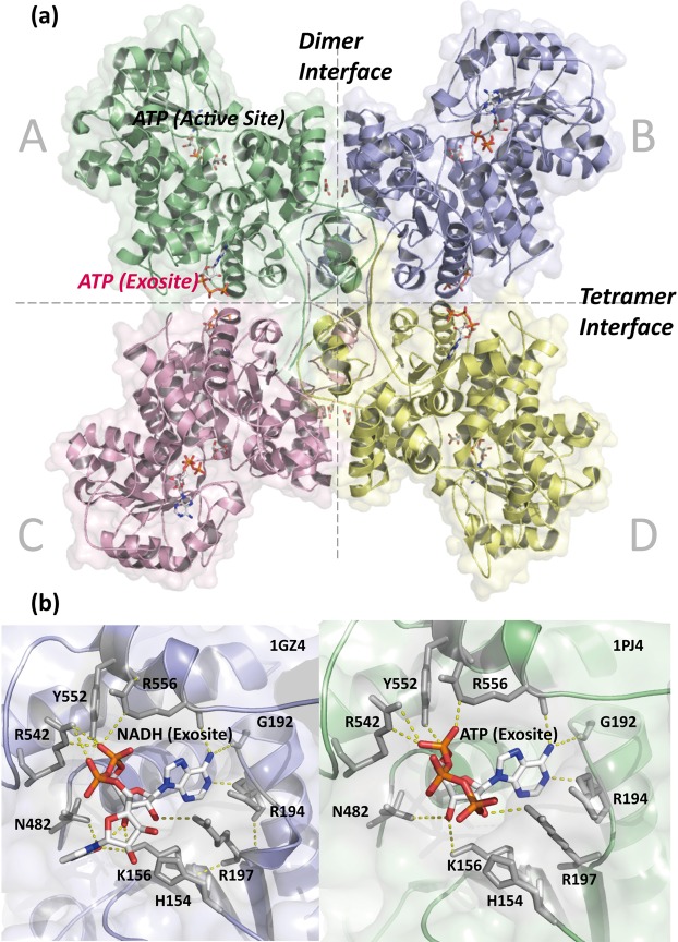
Figure 1.
Structure and sequence alignment of human m-NADP-ME. (a) The crystal structure of human m-NADP-ME shows the dimer interface between the AB or CD dimers, the tetramer interface between the AD or BC dimers and the four active sites and the exosite with ATP in each monomer (PDB code: 1PJ4). (b) The overall binding resides of the exosite, with an NADH-binding mode (PDB code: 1PJ2) and an ATP-binding mode (PDB code: 1GZ4). These figures were generated by using PyMOL41.

