Fig. 3.
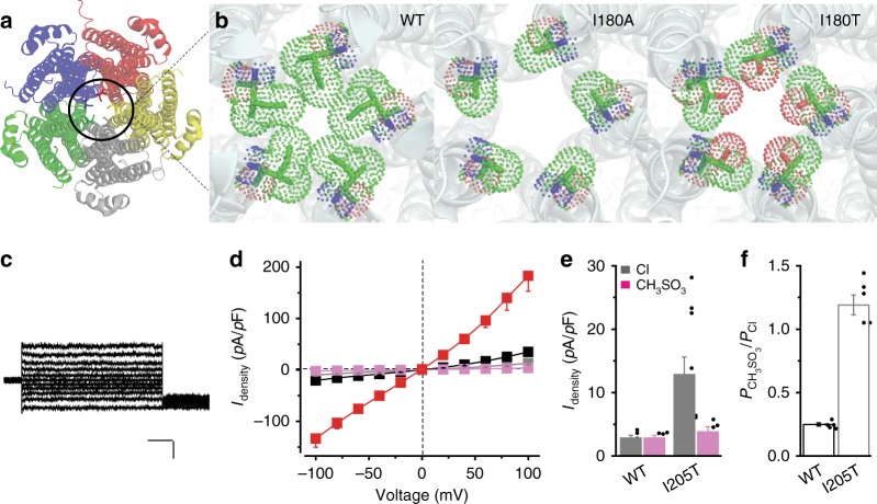
Structural and functional analyses of two aperture mutants. a KpBest pentamer viewed from the cytoplasmic side and vertical to the ion-conducting pathway. b Apertures of KpBest wild-type (WT), I180A, and I180T as viewed from the same direction as in a. Figures were made from actual crystal structures, and critical residues on the apertures are colored by element. c Representative human bestrophin1 (hBest1) I205T current traces in the absence of Ca2+, with Cl− in the external solution. Scale bar, 100 pA, 150 ms. d Population steady-state current density–voltage relationships in HEK293 cells expressing hBest1 I205T, with Cl− in the external solution in the absence (gray) or presence (black) of 1.2 μM Ca2+ or with CH3SO3− in the external solution in the absence (magenta) or presence (red) of 1.2 μM Ca2+, n = 10–11 for each point. e Bar chart showing the steady-state Cl− and CH3SO3− current densities of hBest1 WT and I205T at 100 mV in the absence of Ca2+, n = 5–10 for each bar. f Relative ion permeability ratio (PCH3SO3/PCl) in hBest1 WT and I205T, n = 5–10 for each bar. All error bars in this figure represent s.e.m.

