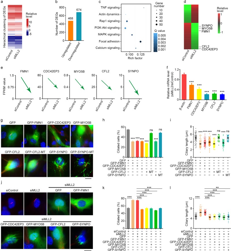
Fig. 2. Downregulation of actin-associated proteins by MLL2 depletion underlies its effect on ciliogenesis.
a, b RPE-1 cells were transfected with control or MLL2 siRNAs and serum-starved for 48 h. The hierarchical clustering of DEGs (a) and number of DEGs (b) were then obtained. c KEGG pathway analysis showing signaling pathways affected by MLL2 depletion. MAPK, mitogen-activated protein kinase; PI3K, phosphoinositide 3-kinase; TNF, tumor necrosis factor. d Heatmap showing the differential expression of proteins by MLL2 depletion. e FPKM value analysis of the relative expression of five actin-associated proteins in RPE-1 cells transfected with control or MLL2 siRNAs and serum-starved for 48 h. f Quantitative RT-PCR analysis of the relative mRNA levels of the indicated proteins upon MLL2 depletion. g–i Immunofluorescence images (g), percentage of ciliated cells (h, n = 100), and ciliary length (i, n = 30) for RPE-1 cells transfected with the indicated plasmids, serum-starved for 48 h, and stained with acetylated α-tubulin antibodies (red) and DAPI (blue). In the DDI/EEV mutant of CFL2 (CFL2-MT), Asp141, Asp142, and Ile143 were replaced by Glu, Glu, and Val, respectively. In the Δ752–903 mutant of SYNPO (SYNPO-MT), amino acids from 752 to 903 were deleted. Scale bar, 10 µm. j–l Immunofluorescence images (j), percentage of ciliated cells (k, n = 100), and ciliary length (l, n = 30) for RPE-1 cells transfected with the indicated siRNAs and/or plasmids, serum-starved for 48 h, and stained with acetylated α-tubulin antibodies (red) and DAPI (blue). Scale bar, 10 µm. **P < 0.01; ***P < 0.001. Error bars indicate SEM

