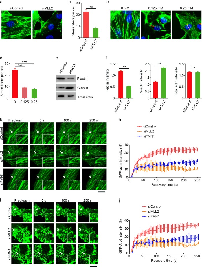
Fig. 3. MLL2 regulates actin dynamics.
a–d Immunofluorescence images (a, c) and quantification of stress fibers (b, d, n = 50) for RPE-1 cells transfected with control or MLL2 siRNAs (a, b) or treated with the indicated concentrations of MTA (c, d), serum-starved for 48 h, and stained with acetylated α-tubulin antibodies (red), FITC-phalloidin (green), and DAPI (blue). Scale bars, 5 µm. e, f Immunoblotting (e) and quantification (f) of F-actin, G-actin, and total actin from RPE-1 cells transfected with control or MLL2 siRNAs and serum-starved for 48 h. g, h RPE-1 cells were transfected with GFP-actin and control, MLL2, or FMN1 siRNAs and serum-starved for 48 h. Images were recorded at 5 s intervals following photobleaching of the indicated area (g) and fluorescence recovery at different time points was quantified (h, n = 10). Insets show higher magnifications of the bleaching regions. Scale bar, 10 µm. i, j RPE-1 cells were transfected with GFP-Arp2 and control, MLL2, or FMN1 siRNAs and serum-starved for 48 h. Images were recorded at 5 s intervals following photobleaching of the indicated area (i) and fluorescence recovery at different time points was quantified (j, n = 10). Insets show higher magnifications of the bleaching regions. Scale bar, 10 µm. **P < 0.01; ***P < 0.001; ns, not significant. Error bars indicate SEM

