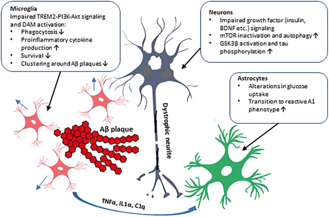
FIGURE 2.

Schematic illustration of the cell type-specific effects of impaired PI3K-Akt signaling in AD brain. The impaired PI3K-Akt signaling might be due to decreased PI3K subunit levels observed in the brain of AD patients or due to T2D, which alters insulin levels and affects PI3K-Akt pathway in the brain, augmenting AD pathogenesis. These alterations could have distinct effects in different cell types in the brain, as shown in the figure. Activation of PI3K-Akt signaling pathway by several extracellular stimuli, such as growth factors (insulin, BDNF, etc.), affects proliferation, metabolism, and survival in various cell types. In neurons, GSK3β activity and tau phosphorylation and mTOR activity affecting autophagy are influenced. Microglial transit to DAM phenotype, phagocytosis, and cytokine production, and formation of microglia clusters around Aβ plaques are affected. Inflammatory cytokines secreted by microglia activate astrocytes, which transit to the reactive A1 phenotype unable to support neuronal outgrowth and synaptogenesis. Furthermore, genetic variation in AKT2 gene alters glucose uptake, probably in astrocytes, potentially affecting brain energy metabolism.
