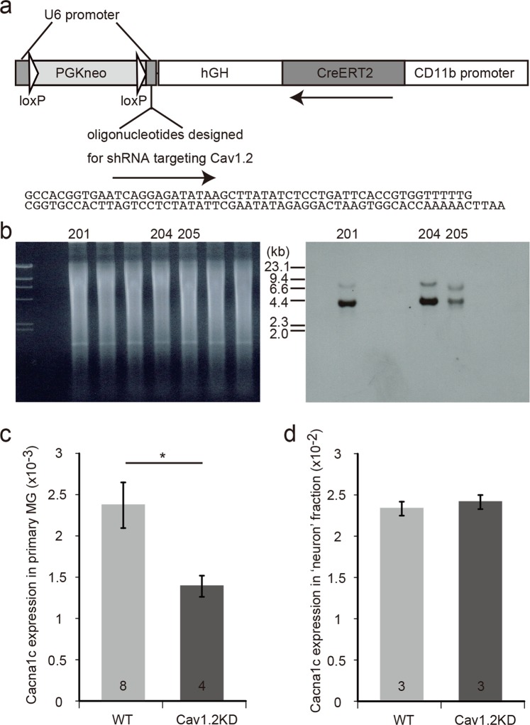
Figure 2.
Generation of Cav1.2KD mice. (a) Schematic structure of the transgene used to generate the Cav1.2KD mice. hGH, 3’ non-coding region of human growth hormone gene. PGKneo, neomycin resistance gene driven by phosphoglycerate kinase promoter. (b) Southern blot analysis to confirm the generation of Cav1.2KD mice. Tail DNA from the F0 founder mice was digested with EcoRI and Southern hybridization was performed using a DIG-labelled CreERT2 probe. The right panel shows the hybridization signals of the blot prepared from the gel shown in the left panel. The expected size of the DNA band derived from the transgene is ~ 4.1 kb. In this experiment, 3 mice were found to be transgenic (No. 201, 204, and 205). The gel and blot presented here are full-length. (c) Cacna1c gene expression in primary microglial cells and ‘neuron’ fraction. Quantification of Cacna1c expression in cultured primary microglia (normalized with GAPDH) by quantitative RT-PCR. Primary cultures were prepared from tamoxifen-treated adult mice of each genotype. The numbers in the columns represent the number of samples, which were obtained from three independent cultures. (d) Cacna1c expression in the ‘neuron’ fraction normalized with GAPDH. The numbers in the columns represent the number of mice used for the preparation of samples. Data are presented as mean ± SEM. *p < 0.05 by Welch’s t-test in (c).

