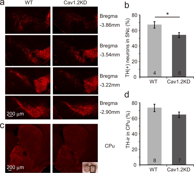
Figure 3.
MPTP-induced degeneration of dopaminergic neurons is enhanced in Cav1.2KD mice. (a) Brain slices containing the SNc area from wild type (WT) and Cav1.2KD mice treated with MPTP were immunostained with a TH-antibody to detect dopaminergic neurons. (b) Number of dopaminergic neurons in the SNc 7 days after MPTP administration, normalized with normal controls (saline treated), was compared between WT and Cav1.2KD. (c) Images of the mouse brain CPu area immunostained with a TH-antibody to show the terminals of the SNc dopaminergic neurons. The boxed area in the inset image represents the CPu area shown in (c). (d) Analysis of the TH-ir signal intensity in the CPu. Scale bars, 200 µm. The numbers in the columns in (b,d) represent the number of mice analysed. Data are presented as mean ± SEM. *p < 0.05 by a Student’s t-test.

