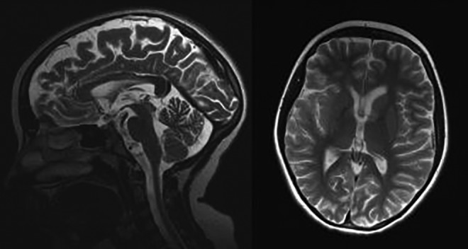
Figure 1.

Brain magnetic resonance imaging (MRI) of the patient performed at the age 10 years. T2-weighted images. A, Abnormal, high signal intensity of the anterior part of the corpus callosum is seen. B, The volume of the basal ganglia is asymmetric and reduced with the hyperintense part of the left head of the caudate nuclei.
