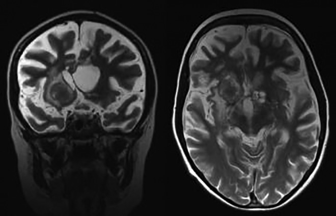
Figure 3.

Magnetic resonance imaging (MRI) examination performed 18 months later, after rituximab administration. T2-weighted images. Focal hyperintense lesion in the right cerebral hemisphere and small cystic lesions on the left side. Note diffuse cerebral atrophy. Progression of lesions.
