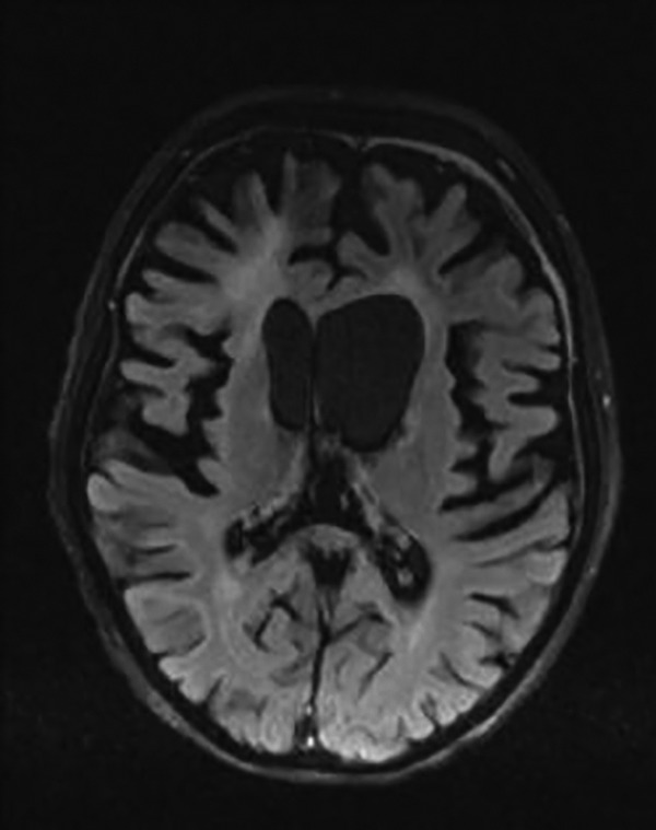
Figure 5.

Brain magnetic resonance imaging (MRI) performed after second chemotherapy course. Axial FLAIR image. Regression of the right focal lesion and the cerebral atrophy in both gray and white matter more prominent in the front parts of the brain. Hyperintense signal of the frontal white matter. Chronic, subdural hematoma in the frontal regions bilaterally.
