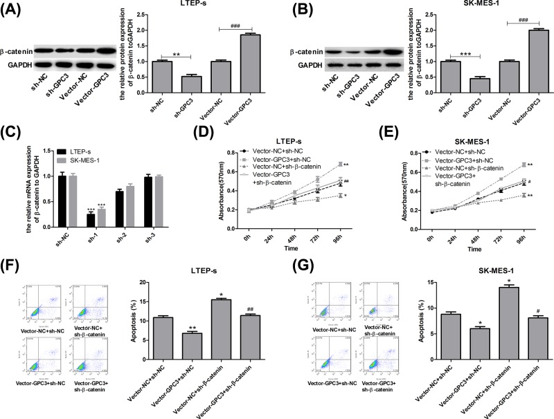
Figure 4. GPC3 promoted cell growth and repressed cell apoptosis through up-regulating β-catenin expression in LTEP-s and SK-MES-1 cells.
(A,B) LTEP-s and SK-MES-1 cells were infected with vector-NC or vector-GPC3, sh-NC or sh-GPC3 for 48 h, and then WB analysis was used to test the expression of β-catenin (n=3, **P<0.01, sh-GPC3 group vs sh-NC group; ###P<0.001, vector-GPC3 group vs vector-NC group). (C) Forty-eight hours after treatments with sh-1, sh-2 and sh-3 targeting the human β-catenin gene and sh-NC, the LTEP-s and SK-MES-1 cells were collected to evaluate the knockdown efficiency by RT-PCR (n=3, ***P<0.001, sh-1 group vs sh-NC group). Then, LTEP-s and SK-MES-1 cells were infected with vector-NC + sh-NC, vector-GPC3 + sh-NC, vector-NC + sh-β-catenin or vector-GPC3 + sh-β-catenin, followed by the following assays. (D,E) An MTT assay was carried out to determine cell proliferation after 0, 24, 48, 72 or 96 h of cell infection. (F,G) Flow cytometry with Annexin V (FITC)/PI staining was carried out to determine cell apoptosis after 48 h of cell infection (n=3, *P<0.05, **P<0.01, ***P<0.001, vector-GPC3 + sh-NC or vector-NC + sh-β-catenin group vs vector-NC + sh-NC group; #P<0.05, ##P<0.01, ###P<0.001, vector-GPC3 + sh-β-catenin group vs vector-GPC3 + sh-NC group).

