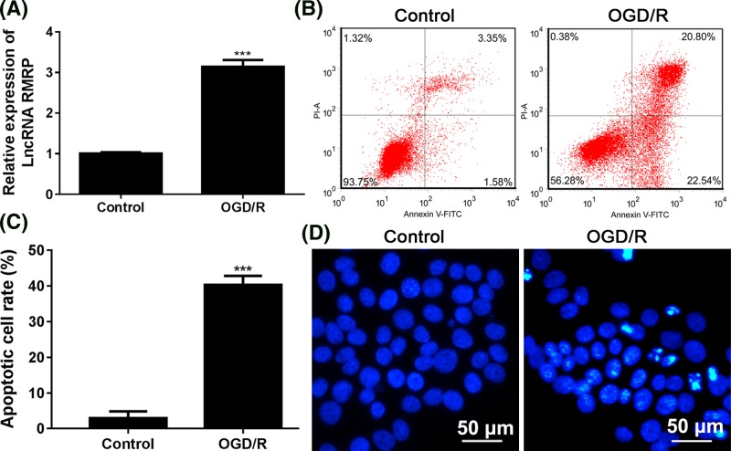
Figure 1. Effect of OGD/R administration on the expression of RMRP and apoptosis in SH-SY5Y cells.
SH-SY5Y cells were cultured in OGD medium in 95% N2 and 5% CO2 for 8 h at 37°C and then the culture was replaced by medium containing 4.5 g/l glucose in an atmosphere of 95% air and 5% CO2 for 24 h. (A) RT-PCR was used to test the result of lncRNA RMRP expression. (B) Apoptotic process and (C) apoptotic cell rates in SH-SY5Y cells were determined using Annexin V/PI staining kit. Flow cytometry was used to detect apoptosis. (D) Hoechst staining was used to test apoptosis. The morphological changes of cell nuclei were detected using a fluorescence microscope at 400× magnification. Apoptotic cells are characterized by pyknotic and fragmented nuclei. After Hoechst 33258 staining, the nucleus of normal cells showed a normal blue color, while the nucleus of apoptotic cells showed somewhat white. ***P<0.001 vs Control group. Each assay was represented by three replicates.

