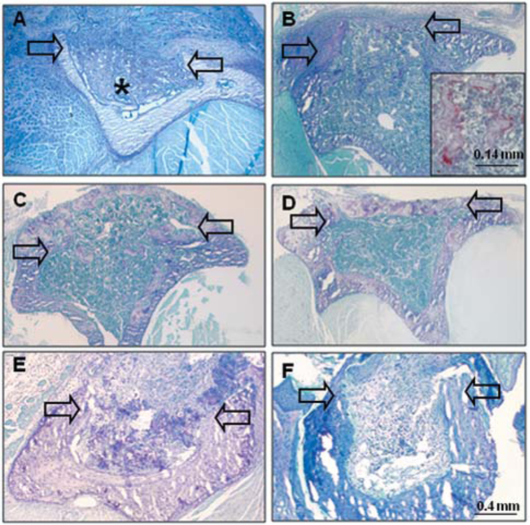
Figure 1.
Histological evaluation of spontaneous repair of tibial wounds in C57BL/6NHsd (A-D), compared with NOD/SCID (E-F) mice. A: At seven days after wounding in C57BL/6HNsd mice, the intramedullary space is filled with reactive woven bone (*). B: At 14 days after wounding, there is woven bone within the wound space and partial repopulation of marrow. Higher power view (inset) shows tartrate- resistant acid phosphatase-positively-stained cells on surfaces of intramedullary bony trabeculae. C: At 21 days after wounding, there is ossification in the cortical wound and repopulation of intramedullary canal with hematopoietic marrow cells. D: At 28 days after wounding, there is bridging of the cortical wound with mature bone. E: At 14 days after wounding in NOD/SCID mice, there is presence in the canal of residual clot and poorly vascularized fibrous tissue, with little evidence of ossification. F: At 28 days after wounding, poorly vascularized fibrous tissue fills the medullary canal. Arrows indicate margins of cortical wound in transverse sections.

