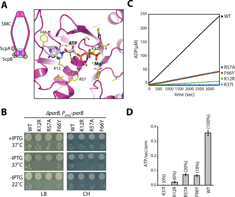
Figure 1. Identification of SMC mutants with decreased ATPase activity.

A, Homology model of the ATPase domain of B. subtilis SMC. Only one of the two composite ATP binding domains is shown. Amino acids that were mutated are indicated (yellow). A schematic of the condensin complex with its head domains (boxed) bound to ATP (blue balls) is shown on the left. B, Growth of wild-type and the relevant mutants in the presence and absence of ParB on agar plates. The 10−2 and 10−5 dilutions are shown. Under Hi-C assay conditions (22˚C no IPTG and 37˚C with IPTG in CH medium), the mutants grow similar to wild-type. C, NADH-coupled ATPase activity assay for the indicated mutants. D, Bar graph showing ATPase activities. Error bars show the standard deviation of four replicates. See also Figures S1, S2, Tables S1 and S2.
