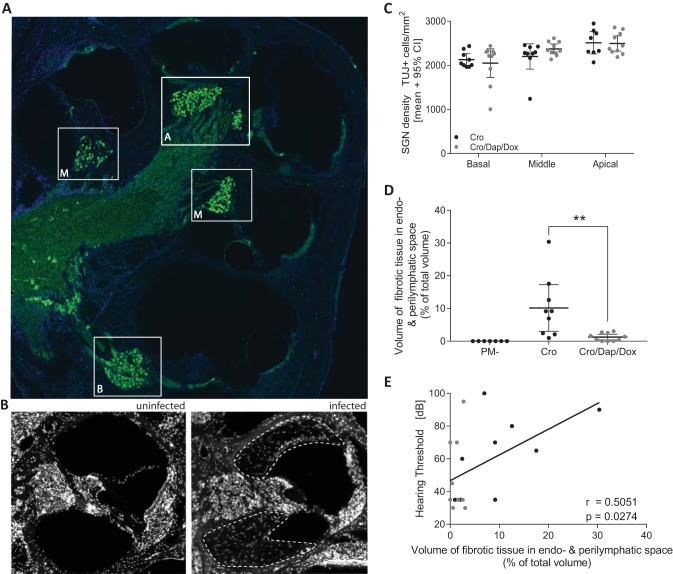
FIG 5.
Immunohistological analysis of spiral ganglion neuron density and cochlear occlusion in mildly infected animals. (A) Midmodiolar section immunostained for neurons (β-III tubulin; green) and cell nuclei (DAPI; blue) showing the basal (B), middle (M), and apical (A) turns of an infected rat 3 weeks after PM. (B) The absence and presence of fibrous occlusion in the perilymphatic space in a mock-infected control animal and in an infected animal receiving standard CRO monotherapy are represented in immunofluorescence pictures. The area with fibrous occlusion in the perilymphatic space is indicated by white dashed lines. (C) Spiral ganglion neuron (SGN) density levels in any of the cochlear turns in infected animals were not significantly different between the two treatment groups. (D) Combined-adjuvant therapy (n = 10) significantly reduced the amount of fibrous tissue in the perilymphatic space compared to that with CRO monotherapy (n = 9). (E) A significant positive correlation for fibrous occlusion of the perilymphatic space and hearing threshold was found. **, P < 0.01.

