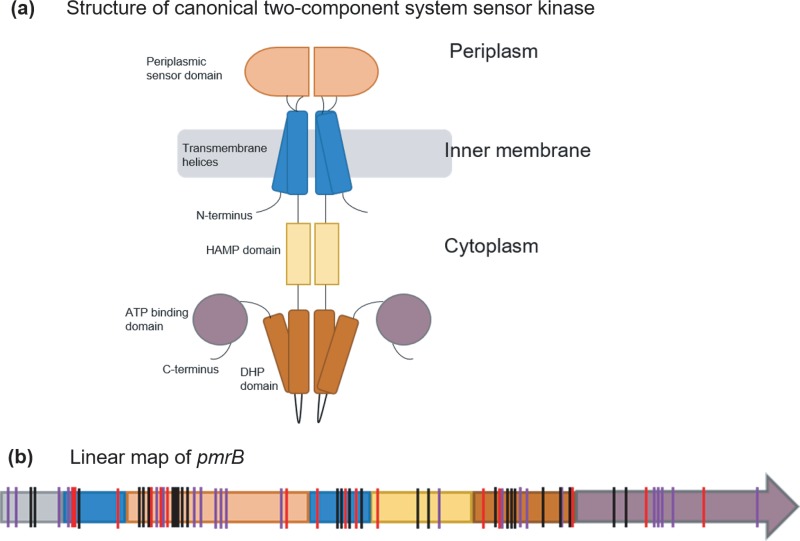
FIG 5.
(a and b) Structural representation of a canonical two-component system sensor kinase dimer (adapted from reference 51 with permission of the publisher) (a) and linear map of pmrB showing positions of identified mutations (b). The color scheme for domains used in panel a is maintained in panel b. (b) Mutations identified in pmrB in this study as well as previous works have been indicated by vertical lines on the pmrB gene. Red lines represent mutations identified in this study. Black lines represent mutations observed in other evolution experiments (12, 13, 28, 32), and purple lines represent mutations identified in colistin-resistant clinical P. aeruginosa isolates (10–12, 27, 33, 34, 52). HAMP, histidine kinases, adenylate cyclases, methyltransferases, and phosphodiesterases; DHP, dimerization and histidine phosphotransfer. Domain assignments in the PmrB protein are based on the predicted domain structure of PmrB by Moskowitz et al. (27). Details of the types of mutations observed in each domain of PmrB in this study can be found in File S1.

