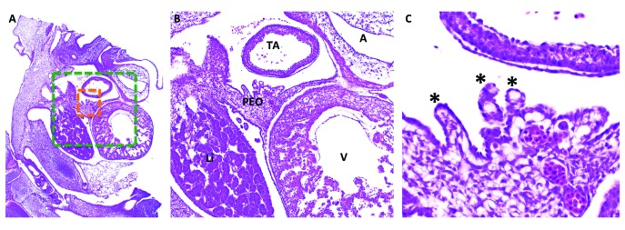
Figure 10.
(A) Histology (hematoxylin and eosin stain) of the developing chick embryo at HH21–23. (B) Green-boxed area in panel A at increased magnification. (C) Orange-boxed area in panel A at increased magnification. The proepicardial organ (PEO) has villous projections (black asterisks) that attach to the myocardial tube and begin to spread over the portion of the tube that will become the ventricle (V), followed by the atria (A) and truncus arteriosus (TA). Li, liver.

