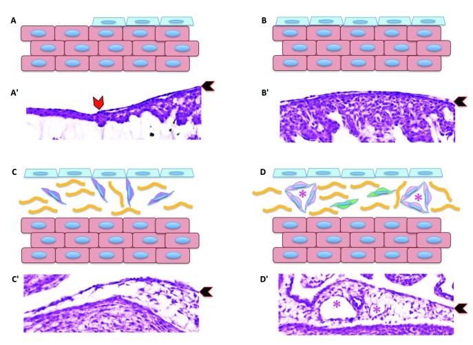
Figure 11.
Schematic diagram showing epicardial migration over the myocardium and the subsequent epithelial-to-mesenchymal transition. (A) The epicardial cell layer (pale-blue cells) migrate over the naked cardiomyocytes (salmon-colored cells). (B) Eventually the entire myocardium is covered by a sheet of epicardial cells. (C) Once the the layer of epicardium covers the entire myocardium, epicardial cells begin the epithelial-to-mesenchymal transition, giving rise to epicardium-derived cells (purple cells). (D) In turn, epicardium-derived cells become cardiac fibroblasts (green cells) or cells contributing to the formation of blood vessels (pink cells), including endothelial cells, smooth muscle cells, and perivascular fibroblasts. Panels Aʹ through Dʹ are corresponding hematoxylin and eosin–stained sections of embryonic chick hearts. Red chevron in panel Aʹ indicates the migrating leading edge of the epicardial cells over the myocardium. Black chevrons in panels Aʹ through Dʹ indicate the epicardium. Yellow lines in panels C and D represent indicate subepicardial connective tissue. Pink asterisks in panels D and Dʹ indicate the blood vessel lumen.

