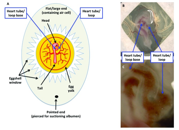
Figure 3.
Ease of visualization and manipulation of the developing chicken embryo. (A) Schematic diagram of a fertilized egg containing a HH16–18 embryo. The end opposite to the air cell has been pierced to remove albumin, and a window in the shell has been created by using jeweler's forceps. (B) Modification of the early chick (EC) culture system, in which chicken embryos within a filter paper frame are transferred to a culture dish. (C) Close-up of panel B.

