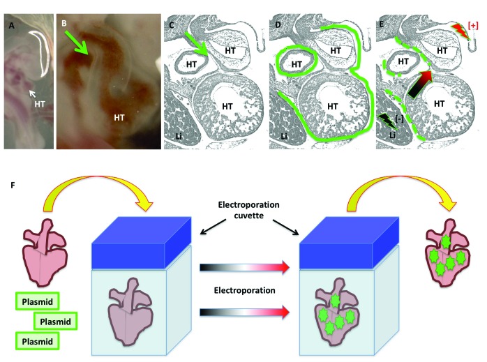
Figure 6.
In ovo and ex ovo epicardial electroporation in embryonic chickens. (A through E) In ovo epicardial electroporation for stages HH21–24. For in ovo epicardial electroporation, (A) the embryo is visualized, and (B and C) the pericardial cavity injected (green arrows) with (D) plasmid solution (green continuous line). Immediately after plasmid injection into the pericardial space, (E) the epicardium is electroporated by delivering currents at the indicated sites (lighting symbols), causing the plasmids to enter the epicardial cells. (F) For ex ovo epicardial electroporation, the heart is dissected and placed inside an electroporation cuvette containing a plasmid solution. Delivery of electric current results in the electroporation of epicardial cells. The green dotted lines in panel E and the green stars in panel F represent the successfully electroporated cells. HT, heart tube; Li, liver.

