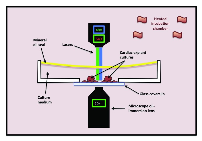
Figure 8.
Set-up for fluorescent imaging of live tissue culture cells. A glass-bottom culture dish containing the fluorescently labeled tissues of interest and culture medium and sealed with mineral oil (to prevent medium evaporation) is placed on the stage of an inverted microscope within a heated incubation chamber. Depicted in this diagram is an inverted fluorescent microscope with a 488-nm laser to visualize the green channel and a 543-nm laser to visualize the red channel at 20× magnification.

