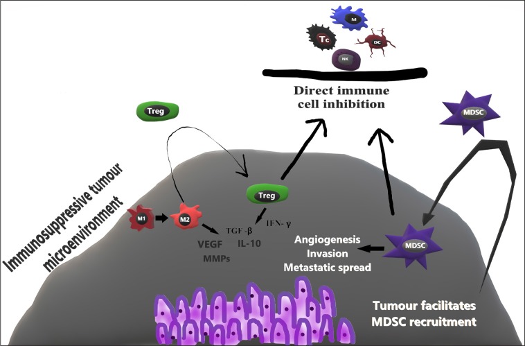
Fig. 3.
Illustration of protumour immune response
NK – natural killer; DC – dendritic cell; M – macrophage; Tc – T cytotoxic lymphocyte; M1–M1 (killer) macrophage; M2–M2 (healer) macrophage; Treg – regulatory T cell; MDSC – myeloid-derived suppressor cell; VEGF – Vascular endothelial growth factor; MMPs – Matrix metalloproteinases; TGF-β – Transforming growth factor beta; IFN-γ – Interferon gamma; IL-10 – Interleukin 10

