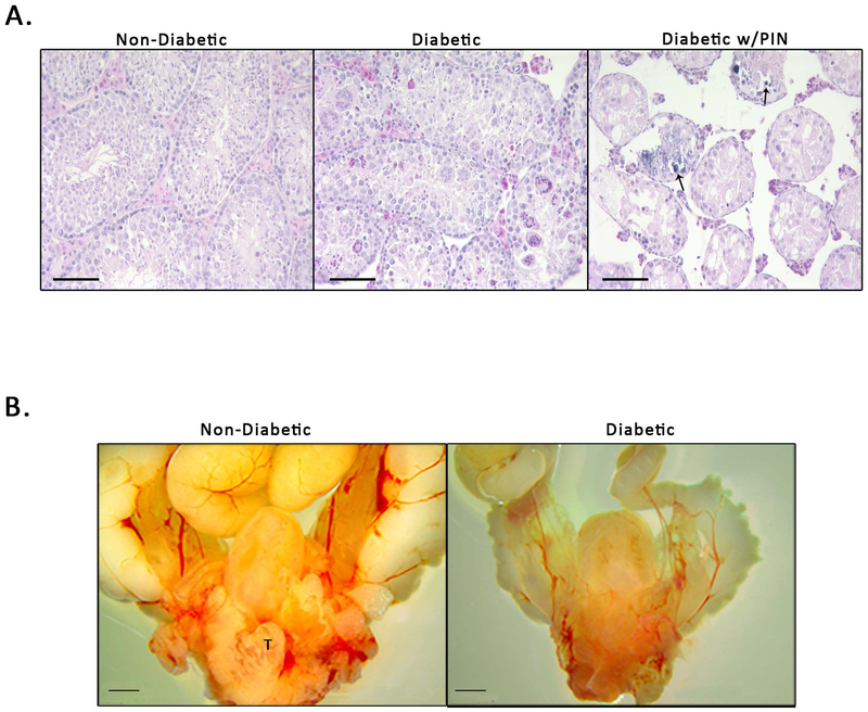
Figure 6. Diabetic Status is Associated with Testicular Changes in the NOD Mouse Prostate.
A) Testicular phenotypes in non-diabetic mice (ND), diabetic mice (Di), and diabetic mice with PIN-like changes (Dp). ND mice appear to have normal phenotypic composition. Di mice exhibit calcification of the testes and cellular atrophy (arrows indicted areas of calcification). Dp mice have complete loss of testicular architecture, loss of Leydig cells, and severe cellular atrophy. B) Gross anatomy shows a decrease in the overall size of the urogenital tract in diabetic NOD mice compared to non-diabetic NOD mice (T indicates visible testis in non-diabetic mouse).

