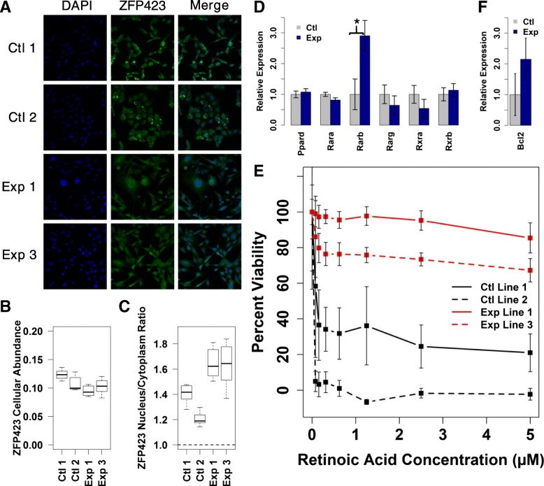
Fig. 6.
Analysis of ZFP423 and retinoic acid effects in SDHC-loss MEFs. a Representative ZFP423 immunostain images of stable SDHC-loss (Exp) and hemizygous control (Ctl) MEF lines. b Analysis of ZFP423 mean cellular immunostain intensity using CellProfiler automated image analysis approach. c Analysis of ZFP423 subcellular localization using CellProfiler automated image analysis. d Relative RNA-seq gene expression quantification for known retinoic acid receptors and transcriptional co-activators. Comparisons indicated by asterisks are statistically significant by a two-sided heteroscedastic t-test (p < 0.05). e Alamar blue cell viability analysis following 6-d exposure of MEF cells to retinoic acid. f Relative RNA-seq gene expression quantification for Bcl2

