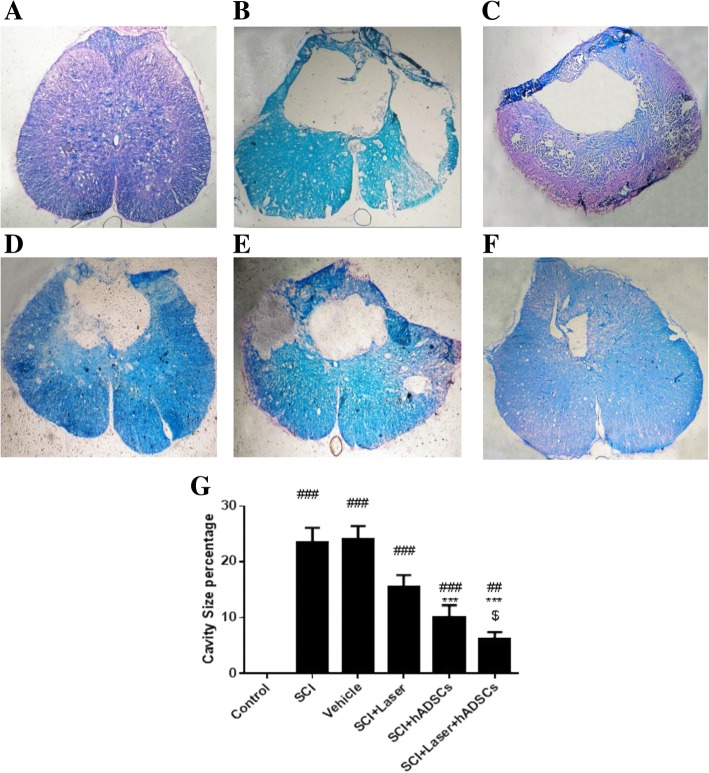
Fig. 6.
Assessment of cavity size and myelinated area by Luxol Fast Blue (LFB) staining 4 weeks after spinal cord injury (SCI), spinal cord transverse section (× 10). The assessment showed that the largest cavity was observed in the SCI group, while smaller cavities were observed in the combination-treated animals. In addition, the most myelinated area was detected in these animals. Control (a); SCI (b); vehicle (c); laser (d); hADSCs (e); laser+hADSCs group (f); quantitative assay of cavity size (g). Data are expressed as mean ± SEM. ##p < 0.01, ###p < 0.001, vs control group, ***p < 0.001 vs. SCI group and vehicle; $p < 0.05 vs laser

