Fig. 1.
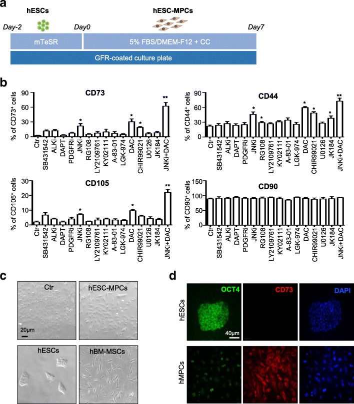
JNKi and DAC initiate generation of hESC-MPCs. a Schematic of mesenchymal differentiation from hESCs to hESC-MPCs. Single H1 hESCs were seeded on GFR-coated 6-well plate for 2 days, then the mTeSR medium was changed into DMEM/F12 media supplied with 5% FBS and/or chemical compound (CC) for a week. b Flow cytometry (FCM) analysis of MSC marker of hESC-derived cells cultured in 5% FBS/DMEM/F12 ± 10 nM chemical compounds for 7 days (mean ± SEM, N = 3). *P < 0.05;**P < 0.01. c Morphology of H1 hESCs cultured in 5% FBS/DMEM/F12 with (hESC-MPCs) or without (Ctr) JNKi and DAC addition for 7 days, undifferentiated hESCs (hESCs), and hBM-MSCs (scale bar = 20 μm). d Representative immunofluorescence images display the expression of OCT4 (in green) and CD73 (in red) in hESCs and hESC-MPCs (scale bar = 40 μm). The nuclei (in blue) were labeled with DAPI

