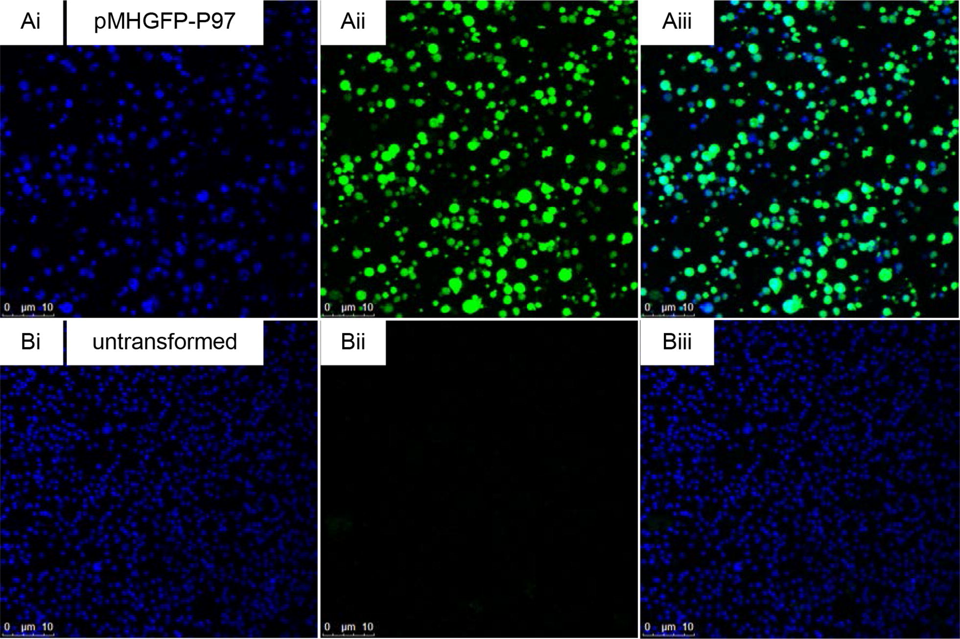
Figure 4.

Confocal micrographs of M. hyopneumoniae (pMHGFP-P97). M. hyopneumoniae cells were stained with 4′,6-diamidino-2-phenylindole (DAPI) and were excited by 405 nm and 488 nm lasers to visualise nucleic acid and green fluorescence, respectively. A (i) M. hyopneumoniae (pMHGFP-P97) cells stained by DAPI, (ii) M. hyopneumoniae (pMHGFP-P97) cells viewed on green fluorescence channel, (iii) overlay of i and ii. B (i) untransformed M. hyopneumoniae cells stained by DAPI, (ii) untransformed M. hyopneumoniae cells viewed on green fluorescence channel, (iii) overlay of i and ii.
