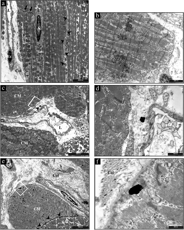
Fig. 7.
TEM analysis of the heart from a representative untreated CTRL and TiO2-NP treated SHR. a low magnification image of a CTRL heart to illustrate the sarcolemma (arrowheads) lining the surface and gap junctions (arrows) delimiting cardiomyocytes filled of mitochondria and myofibrils. On the left, collagen bundles (Col) are present in the interstitial space between an endothelial cell (Ec) lining a capillary lumen (L) and cardiomyocytes. N: cardiomyocyte nucleus. b aggregates of TiO2-NPs within the cardiomyocyte cytoplasm showing effaced myofibrils and swollen mitochondria. c the white rectangle inscribes an area shown at higher magnification in (d) in which the arrowhead points to NP located in the interstitial space between the vessel wall inscribing a lumen (L) and cardiomyocytes (CM). e low magnification image of a treated SHR myocardium to illustrate widening of the interstitial space by abundant fibrotic deposition (Col) surrounding cardiomyocytes (CM) and an endothelial cell (Ec) lining a capillary. Arrowheads point to gap junctions delimiting two CMs. The white rectangle inscribes an area shown at higher magnification in f to document the internalization of TiO2-NP located in proximity of the CM sarcolemma bordered by collagen bundles (Col). Scale Bars: A, C, E: 5 μm, B: 2 μm, D: 1 μm, F: 500 nm

