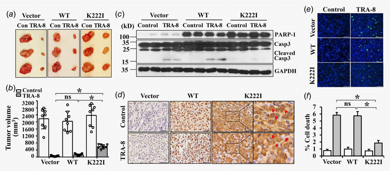
Figure 3.
Cytoplasmic PARP-1 increases resistance of sensitive pancreatic cancer to TRA-8 therapy in mice. BxPc-3 cells stably infected with Vector, wild-type PARP-1 (WT) or the PARP-1 cytoplasmic mutant (K222I) were injected into nude mice, which were then subjected to control vehicle (Control, Con) or TRA-8 treatment for 6 weeks. (a) Representative tumors; and (b) Tumor volumes in each group are shown (n = 8–9/ group, ns = not significant, *p < 0.01). (c) Western blot analysis of the expression of PARP-1 and activation of caspsase-3 (Casp3) in representative tumors in each group. The expression of GAPDH was used as a loading control. (d) Immunohistochemical staining of PARP-1 in representative tumor sections from each group (Scale bar = 50 μm). Higher magnification of the boxed area are shown to the right. Red arrows indicate cytoplasmic localization of PARP-1 in tumors with the K222I PARP-1 mutants. (e and f) Cell death in tumors from each group was analyzed by TUNEL staining. (e) Representative TUNEL staining images from each group (Scale bar = 50 μm). (f) Quantitative analysis of TUNEL positive cells as percentage of total cells in the tumor sections (n = 5, *p < 0.01).

