
Fig. 2 |. Domain organization of proteins implicated in polycystic kidney disease.
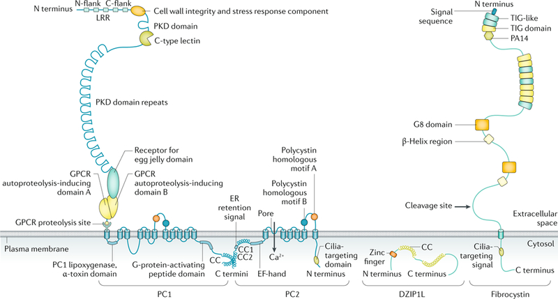
The structure of polycystin 1 (PC1), polycystin 2 (PC2), the membrane-bound form of fibrocystin and DAZ-interacting protein 1-like protein (DZIP1L) are depicted (not to scale). PC1 and PC2 are multispan membrane proteins that form a complex that is localized to multiple subcellular locations, including the primary cilium. Fibrocystin is also localized to the primary cilium and is subject to Notch-like proteolytic processing, resulting in release of the carboxy-terminal tail, which can translocate to the nucleus and may regulate gene expression. DZIP1L is a soluble zinc-finger protein that is localized to the centrioles and basal bodies at the ciliary transition zone. CC, coiled coil; C flank, carboxyl flank; C terminus, carboxyl terminus; ER, endoplasmic reticulum; GPCR, G protein-coupled receptor; LRR, leucine-rich repeat; N flank, amino flank; N terminus, amino terminus; PKD, polycystic kidney disease. Adapted from REF.293, Springer Nature Limited.