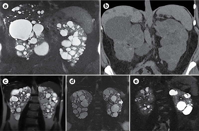
Fig. 7 |. Diagnosis of autosomal dominant polycystic kidney disease using different imaging techniques.

a–b | Comparison of MRI (T2; part a) and CT (without contrast; part b) scans of a 35-year-old woman with autosomal dominant polycystic kidney disease (ADPKD), showing widespread kidney cysts and a few liver cysts. c–e | T2 MRI images of patients with ADPKD who have a truncating mutation in PKD1 (part c; 41-year-old man), a non-truncating mutation in PKD1 (part d, 40-year-old man) or a splicing mutation in PKD2 (part e; 41-year-old man). Patients with truncating mutations in PKD1 typically have more cysts, whereas patients with non-truncating mutations in PKD1 have an intermediate number of cysts and patients with splicing mutations in PKD2 have the fewest cysts.
